
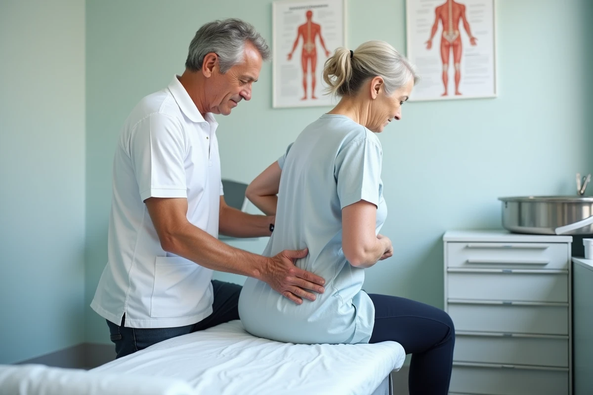
Treating a pressure ulcer on the back involves precisely identifying the pressure area in question, the stage of the lesion, and the aggravating factors specific to the patient. The dorsal location, from the sacrum to the shoulder blades, concentrates specific mechanical constraints that simple rotation in bed does not always resolve. What positioning parameters, local care, and devices truly make a difference in the healing of a dorsal pressure ulcer?
Semi-Fowler Position 30° and Repositioning Plan for Dorsal Pressure Ulcers
Most articles on pressure ulcers mention the necessity of changing position regularly. What is less often detailed is the precise angle of the torso and its direct impact on the pressure exerted on the sacrum and lower back.
See also : How to Reset a Parkside Robotic Mower: Tips and Essential Steps
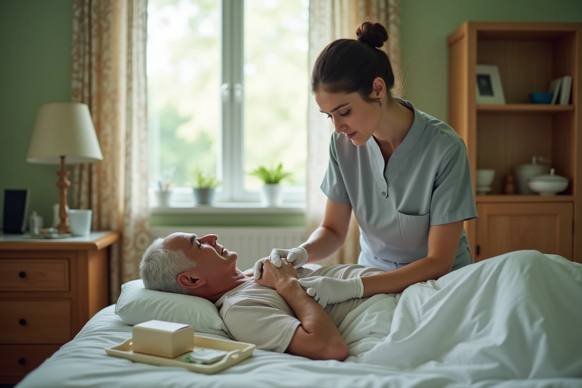
Recent nursing care guidelines recommend semi-Fowler position at 30° rather than flat dorsal position. This slight incline of the torso reduces pressure on the sacrum, lower back, and heels. The recommended alternation is as follows: dorsal position 30° and lateral position 30° every 2 to 3 hours, with an individualized positioning plan documented in the care record.
To learn how to treat a pressure ulcer on the back, this angular data is a concrete starting point, much more effective than a simple reminder to mobilize every two hours.
Related reading : How to Access the Digital Class Paris: A Practical Guide for Students
Skin sliding is a common trap in the semi-sitting position. When the patient slides down the bed, the tissues of the back and sacrum experience shear forces that worsen the existing wound or create a new one. Multi-position positioning cushions, sometimes filled with microbeads, help stabilize the body at 30° and limit this sliding.
Pressure Ulcer on the Back: Comparative Table of Local Care by Stage
The local treatment of a dorsal pressure ulcer directly depends on the stage of the lesion. The actions differ when facing persistent redness versus a wound exposing deep tissues.
| Stage | Skin Appearance | Recommended Local Care | Primary Objective |
|---|---|---|---|
| Stage 1 | Persistent redness, intact skin | Immediate offloading, gentle stroking with flat hand, no firm massage | Restore local vascularization |
| Stage 2 | Blister or superficial skin loss | Hydrocolloid or hydrocellular dressings, cleaning with saline solution | Maintain a moist environment conducive to healing |
| Stage 3 | Tissue loss reaching subcutaneous tissue | Absorbent dressings suitable for the level of exudate, debridement if necrosis | Control infection, promote granulation |
| Stage 4 | Deep tissue involvement (muscle, bone) | Specialized care, possible surgical debridement, complex dressings | Avoid superinfection, prepare the wound bed |
A often underestimated point: the High Authority of Health has prohibited massaging reddened areas for over twenty years. The recommended practice remains gentle stroking with a flat hand, without pressure, combined with immediate offloading in case of persistent redness. Massaging an early-stage pressure ulcer worsens the destruction of weakened tissues.
Management of Exudate and Dressing Selection
At stages 2 and 3, the choice of dressing depends on the volume of exudate. A dressing that is too absorbent dries out the wound bed and slows healing. Conversely, an insufficiently absorbent dressing causes peripheral maceration that enlarges the injured area.
The evaluation must be reassessed at each dressing change. An appropriate dressing maintains a moist environment without excess, a documented condition for optimal healing of chronic wounds.
Prevention of Dorsal Pressure Ulcers: Aggravating Factors to Monitor
Repositioning and local care only work if systemic factors are corrected in parallel. Three levers have a direct impact on the risk of pressure ulcers on the back and the speed of healing of an existing lesion:
- Malnutrition: insufficient protein intake slows tissue repair. In long-term bedridden patients, regular nutritional assessment conditions the response to local treatment.
- Skin maceration: prolonged moisture (incontinence, excessive sweating) weakens the skin barrier of the back and sacrum. The use of appropriate absorbent protections and barrier creams limits this factor.
- Prolonged immobility without a positioning plan: without a written protocol and monitoring, position changes are irregular. A plan documented in the care record, with schedules and positions, significantly reduces the risk of recurrence.
Multi-Position Positioning Cushions: A Specific Medical Device
Modular devices designed for offloading dorsal and lumbar areas are now offered as full-fledged medical devices. These cushions can be placed under the knees, ankles, back, or arms depending on the adopted position.
Their benefit lies in stabilizing the position at 30° and reducing shear forces. An air mattress or memory foam does not replace these positioning supports: both act on complementary mechanisms (overall pressure distribution for the mattress, angular maintenance for the cushion).
Healing a Pressure Ulcer on the Back: How the Stage Affects Treatment Duration
The stage at the time of diagnosis largely determines the care trajectory. A stage 1 pressure ulcer detected and offloaded quickly can regress in a few days. A stage 3 or 4 pressure ulcer may require several months of care, with a risk of infectious complications that further prolongs the process.
Early detection remains the most significant factor affecting healing duration. Persistent redness on the back after thirty minutes of offloading warrants immediate nursing assessment.
The management of a dorsal pressure ulcer is never solely local. It combines precise positioning (semi-Fowler 30°, scheduled alternation), wound care appropriate to the stage, correction of systemic factors, and medical positioning devices. The individualized repositioning plan documented in the care record remains the pivotal document coordinating all these interventions on a daily basis.